Bartholin's Gland
Disease: Surgical
Treatment Options
Advanced CO2 laser ablation and nerve-sparing gland excision, performed by a double board-certified Urogynecologist who specializes in complex pelvic anatomy and recurrent cases after prior procedures.
Understanding Bartholin's Gland Disease
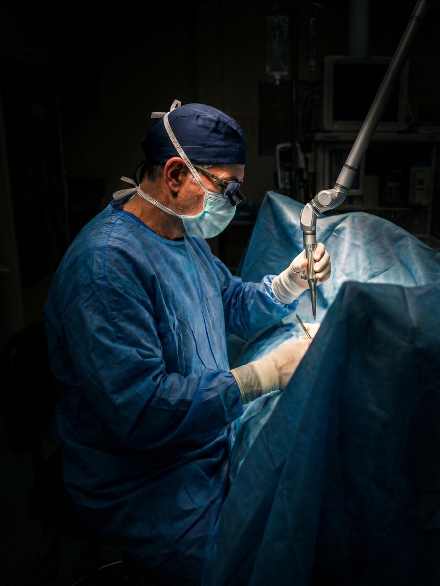
The gland is not hollow. This is the foundational misunderstanding that drives suboptimal surgical decision-making. The Bartholin's gland is a solid, compound tubuloalveolar structure — architecturally comparable to a raspberry, composed of multiple small secretory acini clustered around a branching ductal tree. It is not a balloon, a sac, or a hollow cavity. It does not empty like a cyst. These glands produce the vast majority of their secretion in response to sexual arousal; under normal resting conditions they are relatively quiet, maintaining only a minimal baseline of moisture sufficient to keep the duct pathways clear. The secretions drain through progressively larger collecting ducts and ultimately exit through the main Bartholin's duct at the 4 o'clock and 8 o'clock positions of the vestibular opening.
The cyst forms in the duct — not in the gland. When the main duct becomes obstructed — through inflammation, trauma, inspissated secretion, or scarring from prior procedures — the secretions produced by the intact glandular acini behind the blockage have nowhere to go. The duct itself dilates under the accumulating pressure, forming what is clinically recognized as a Bartholin's duct cyst. The solid glandular tissue posterior to the obstruction remains entirely intact, continues to function, and continues to secrete. This distinction is not semantic; it is the key to understanding recurrence.
Two distinct cyst subtypes — and why the distinction matters surgically. In greater than 90% of cases, the Bartholin's cyst forms entirely within the duct, with the solid glandular tissue remaining uninvolved. In a smaller but clinically significant subset — more common in patients with multiple prior surgeries, particularly those in whom the duct was partially or completely removed during a previous procedure — the cyst will involve the gland itself, or arise from within it. This distinction is consequential: conservative ablation can only address a cyst whose interior lining is accessible and identifiable. If the cyst involves the acinar gland tissue, the laser or RF probe cannot effectively ablate a complex, irregular glandular cavity — and the result will predictably fail. Gland involvement must be suspected in any patient with a history of operative procedures on the Bartholin's apparatus, and excision rather than ablation should be planned accordingly.
When does removing the cyst without the gland cause recurrence? The answer is conditional, and this precision matters. If a surgeon opens or drains the cyst while preserving a functional, patent duct, the gland can continue draining normally and recurrence risk is lower. The problem arises when the duct is removed along with the cyst — an approach that eliminates the gland's only drainage channel. With the duct absent, the intact acini continue secreting, the secretions accumulate, and a new cyst system is inevitable. This scenario is particularly common in patients who underwent a procedure described as "cyst removal" by a surgeon unfamiliar with ductal anatomy: the duct was excised, the gland was left, and recurrence followed as a predictable consequence of the anatomy.
The surgical anatomy surrounding the gland is formidable. The Bartholin's gland occupies a surgically demanding anatomic neighborhood. Immediately adjacent structures include the bulbospongiosus muscle, the crus of the clitoris, the vestibular venous plexus, the pudendal nerve, and — posteriorly — the anal sphincter and rectum. This proximity is precisely why general gynecologists appropriately decline these cases: without regular operative familiarity with this territory, inadvertent injury to any of these structures carries significant functional consequences. In the hands of a surgeon trained specifically in pelvic floor reconstruction, however, these planes are reliably defined, the neurovascular structures are identified and protected, and the procedure is completed safely.
About the Surgeon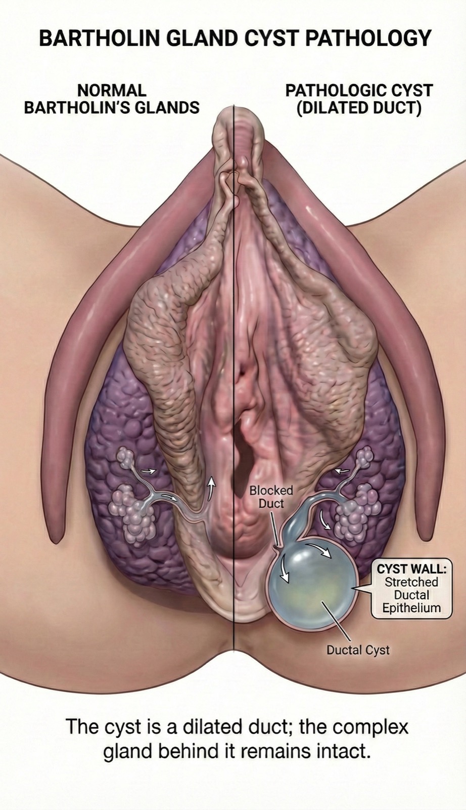
The Bartholin's cyst is a dilated duct. The solid glandular tissue posterior to the obstruction remains intact and continues to secrete — the critical architectural fact that drives recurrence when the gland is not addressed.
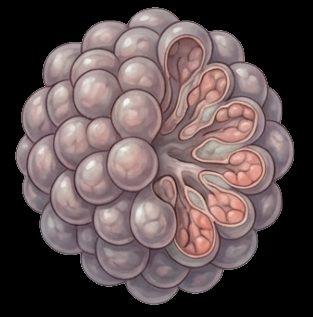
Cross-section of the Bartholin's gland, illustrating its solid, multi-acinar architecture. This is not a hollow sac — it is a compound secretory gland that produces mucoid secretion primarily in response to sexual arousal. The cyst forms upstream in the duct, not within this tissue.
Why Recurrence Follows Standard Management
Understanding the anatomical limitations of conventional approaches is essential to appreciating why specialist intervention is indicated for recurrent disease.
Incision and Drainage (I&D)
Emergency department I&D decompresses the abscess or cyst but does not restore ductal patency or address the glandular architecture. The incision is frequently placed on the thinnest overlying skin rather than at the native duct orifice. If the duct remains obstructed after drainage — which it typically does — the intact glandular acini continue producing secretion, the blockage persists, and a new cyst accumulates. Recurrence rates following simple I&D range from 30% to 50% in the published literature. Only when the procedure incidentally re-establishes a functional drainage pathway does the recurrence risk decrease, and this is not the goal or the typical result of emergency drainage.
Word Catheter Placement
The Word catheter is designed to create a fistulous tract through passive epithelialization over 4–6 weeks. Its efficacy depends on precise placement at the duct orifice and catheter retention for the full dwell period. Premature displacement — which is common — prevents formation of a mature, patent duct and results in recurrence.
Marsupialization
Marsupialization creates a surgically constructed opening by opening the cyst and suturing its everted edges to the vestibular skin. Normal wound healing can re-epithelialize and narrow that opening over time. Careful technique, wound care, and follow-up are important, but no adjunct has been established to reliably prevent recurrence.
The Role of Anatomic Expertise
Bartholin's gland disease affects approximately 2% of women, and the majority of cases encountered by a general gynecologist are managed with I&D or Word catheter placement. Most general gynecologists see only a few Bartholin's cases annually, and very few of those involve complex excision. Surgeons without advanced pelvic floor training may appropriately decline excision given the proximity of the bulbospongiosus musculature, pudendal neurovascular structures, and vestibular venous plexus — anatomic territory that falls outside routine gynecologic training.
The Urogynecologic Advantage
Dr. Stepp's dual fellowship training in Urogynecology and MIGS provides weekly operative experience with precisely the anatomic structures that make Bartholin's gland excision challenging: the bulbospongiosus muscle, the crus of the clitoris, the vestibular venous plexus, the pudendal nerve, and the anal sphincter and rectum. A surgeon who works in this territory up to 4 to 6 times per week understands the dissection planes intimately, protects the surrounding structures reliably, and completes the excision safely. The complexity is real; the risk is manageable with the appropriate training.
The Reconstructive Imperative
Multiple failed drainage procedures frequently leave residual scarring, asymmetry, and altered vulvar architecture. Surgical management should therefore incorporate reconstructive principles: precise tissue plane dissection, meticulous hemostasis, and attention to cosmetic outcome — restoring both function and anatomic symmetry where possible.
A Spectrum of Anatomically Tailored Care
The appropriate intervention is determined by cyst size, recurrence history, degree of scarring, prior procedures, and patient goals. A single surgical approach does not suit all presentations.
CO2 Laser & RF Ablation
Ablation is appropriate when the cyst is confined to the duct — the anatomy present in over 90% of presentations. CO2 laser energy and radiofrequency (RF) ablation both vaporize the internal lining of the dilated duct; as the tissue heals, it creates a lasting drainage channel while leaving the intact acinar gland architecture undisturbed. The gland continues to secrete normally through the newly patent pathway.
While most published studies utilize CO2 laser, Dr. Stepp uses both modalities and selects between them based on anatomy. CO2 laser delivers a straight column of energy — effective for straightforward cysts with a clear linear duct axis. Radiofrequency ablation, by contrast, delivers energy that can be directed around curves, making it the preferred choice for deep cysts, anatomically complex presentations, or cases where the cyst geometry is not linear. The ability to choose the right energy modality for each individual anatomy is a meaningful clinical distinction.
Ablation is most effective when the duct cyst is adequately formed — approximately grape-sized or larger — to permit complete treatment of the duct lining. It is not appropriate when the cyst involves the gland itself, as the irregular acinar cavity cannot be effectively ablated. Gland involvement should be suspected in patients with prior cyst-removal surgery.
Nerve-Sparing Gland Excision
Excision is indicated when ablation is not appropriate or has failed: in cases of gland involvement, deeply scarred anatomy, or presentations where a prior surgery removed the duct but left the secreting gland behind. When the duct is absent and the gland remains intact, recurrence is a near-certainty — the acini will continue producing secretion that has no exit pathway. Removing the gland unit eliminates the secretory source entirely.
The surgical challenge is the anatomy: the bulbospongiosus muscle, the crus of the clitoris, the vestibular venous plexus, the pudendal nerve, and the posterior proximity of the anal sphincter and rectum all require meticulous, plane-by-plane dissection. Dr. Stepp's fellowship training in precisely this anatomic territory — operating in it up to 4 to 6 times per week — makes nerve-sparing gland excision safe and predictable at this practice. An active cyst is not required; excision may be scheduled at the patient's convenience.
Vulvar Reconstruction
Multiple failed drainage procedures can produce vulvar scarring, asymmetric labial architecture, and altered vestibular anatomy. As a board-certified reconstructive pelvic surgeon, Dr. Stepp addresses this residual damage with careful tissue-plane dissection, layered closure, attention to symmetry, and close postoperative follow-up. Reconstructive repair may be performed concurrently with gland excision.
Procedural Approach & Patient Logistics
Anesthesia Options
CO2 laser ablation may be performed under local anesthesia in the clinic setting for appropriate candidates. Nerve-sparing excision is performed under IV sedation or general anesthesia in an outpatient surgical center. Both options are available on-site. Patients receiving sedation must have a responsible adult companion for transport. Post-operative pain is managed with a standardized protocol; most patients describe discomfort as mild to moderate and well-controlled by the first post-operative day.
Optional Post-Operative Adjuncts
Surgical technique, careful wound care, and close follow-up remain the foundation of treatment. If an optional adjunct is discussed, it is identified by name and reviewed for its purpose, evidence, alternatives, risks, and regulatory status. It is not a substitute for complete excision or reconstructive technique.
Travel Coordination for Out-of-Area Patients
Advanced Pelvic Surgery and Urogynecology of Charlotte evaluates complex Bartholin's disease for local and out-of-state patients. Travel patients follow a structured four-step protocol to minimize total travel time and ensure appropriate post-operative monitoring prior to return travel.
- Step 1 — Virtual Records Review: Comprehensive review of prior operative reports, pathology, and imaging. A virtual consultation is conducted to confirm candidacy and establish a surgical plan.
- Step 2 — Arrival & Preparation: The office is easily accessible from Charlotte Douglas International Airport (CLT), with multiple hotels within a 10-minute radius of the practice. Prescription medications may be obtained at the pharmacy directly across from the practice the day before surgery.
- Step 3 — Outpatient Procedure: Performed under appropriate anesthesia at the outpatient surgical center or in-office, depending on the procedure selected. Most procedures are completed within 60 to 90 minutes. A responsible adult companion is required for same-day discharge whenever sedation or anesthesia is provided.
- Step 4 — 24-Hour Local Observation: Out-of-area patients are asked to remain locally for 24 hours following surgery. Most patients receive clearance to return home by commercial air the following day. Post-operative follow-up may be conducted virtually.
Clinical Questions & Answers
Questions addressed here reflect those most commonly raised during consultation and are provided for educational reference only. They do not constitute individualized medical advice.
Out-of-Network by Design
Advanced Pelvic Surgery and Urogynecology of Charlotte operates as a concierge, out-of-network surgical practice with no monthly membership fees. This model allocates the consultation time, operative planning, and direct post-operative access that complex Bartholin's cases require.
Financial Transparency
Upfront procedure pricing is provided prior to scheduling. Payment is due at the time of service. Detailed superbill documentation and procedure coding letters are provided for patients to submit for out-of-network reimbursement from their insurance carrier.
Expedited Scheduling
Complex or urgent Bartholin's presentations are triaged for priority scheduling. Patients with an active abscess or rapidly enlarging cyst are accommodated as soon as logistically possible, bypassing standard wait queues.
Direct Surgeon Access
All surgical patients receive Dr. Stepp's personal contact information for direct post-operative communication. Questions and concerns following surgery are addressed directly with the operating surgeon, not through a call center or on-call service.
Physician Referral Program
Referring physicians receive a detailed operative summary and post-operative care coordination. Dr. Stepp is available for direct physician-to-physician consultation on complex presentations prior to patient transfer of care.
I look forward to helping you understand the right surgical path. — Kevin Stepp, MD, FACOG, URPS