OHVIRA & Vaginal Septum Treatment
An OHVIRA diagnosis can bring questions about pain, surgery, and the future. We can help you understand your anatomy in plain language and discuss the next steps together.
Personal Care Through Evaluation and Treatment
A diagnosis like this often arrives after weeks or months of pain and unanswered questions. APSU is organized so that the time, explanation, and personal attention you need are part of the practice itself.
Time to be heard
Dr. Stepp built this practice to stay small, so appointments leave room to listen, draw out your anatomy, and talk through what treatment can and cannot do. You should leave every visit understanding your own body better than when you arrived.
A concierge-style practice
APSU is organized around personal contact and attentive coordination, so your questions have a clear place to go. The way the practice works is described in full on our Concierge Care page.
What Is OHVIRA?
OHVIRA stands for Obstructed Hemivagina and Ipsilateral Renal Anomaly. It is a condition a girl is born with, arising before birth when the structures that form the uterus, vagina, and kidneys develop along a slightly different path. It was formerly known as Herlyn-Werner-Wunderlich syndrome. The name describes the three things typically found together:
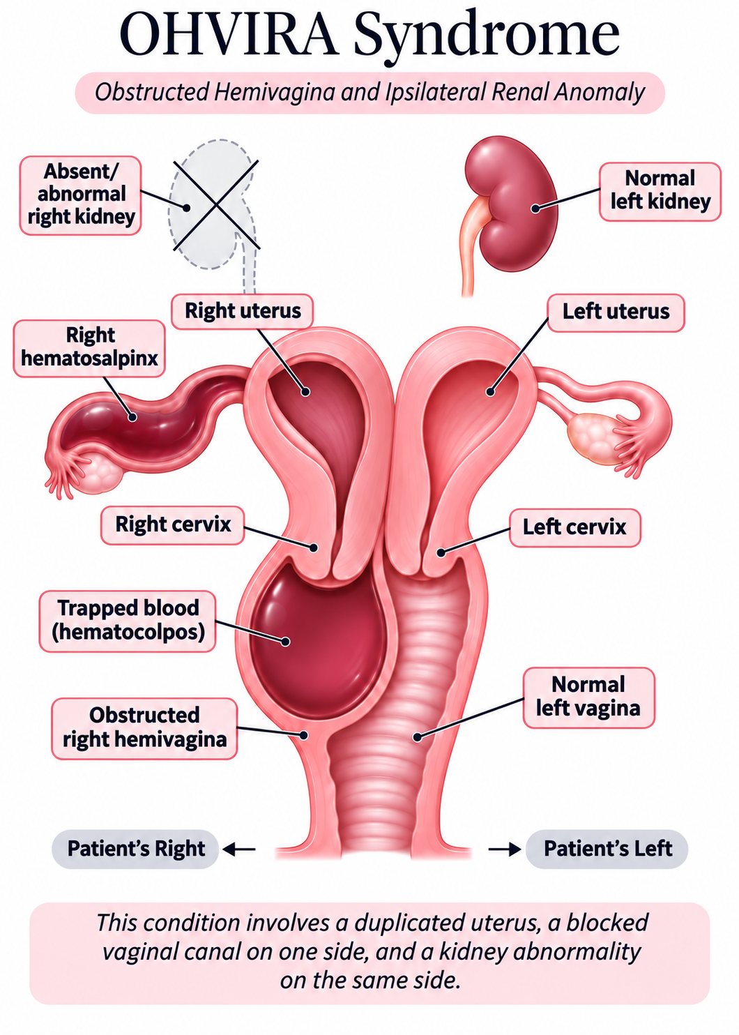
- Duplication of the uterus — most often uterus didelphys, in which the uterus forms as two separate horns, each with its own cervix.
- A vaginal septum with one blocked side — a wall divides the vagina lengthwise, and one side (the hemivagina) has no opening. Menstrual blood from that side’s uterus has nowhere to go.
- A kidney difference on the same side — classically the kidney on the side of the blockage did not develop (renal agenesis). The other kidney usually carries the body’s filtering work.
The trapped flow is what causes the symptoms. After a girl starts her periods, one side bleeds normally while the other side fills and stretches the blocked hemivagina. Doctors call this collection hematocolpos, and it can back up further into the uterus (hematometra) or fallopian tube (hematosalpinx). The result is often monthly cramping that worsens over time, pelvic pain between periods, or a pressure sensation — even though periods may look regular because the open side bleeds normally. That combination, normal periods plus worsening pain, is one of the reasons the diagnosis so often takes a while to reach.
Every case is its own anatomy. The septum can be high or low, partial or complete. The degree of duplication varies, and the kidney finding can range from an absent kidney to a kidney that formed differently. OHVIRA sits within a wider family of congenital uterine and vaginal differences — including uterus didelphys, bicornuate uterus, and vaginal septa of different kinds — and each is its own pattern. This is why careful imaging and an individualized plan matter more than any single standard answer. Published reviews describe OHVIRA as rare, with recognition often delayed by years; a 2025 meta-analysis of the published literature estimated average age at symptom onset around 14 and average age at diagnosis after 16. Getting a clear diagnosis later is common and is a reflection of how rarely this condition appears, and there is real value in bringing a suspected diagnosis forward for proper evaluation now.
Understanding the Anatomy Helps
Here is the classic pattern described in the medical literature, step by step. Real anatomy varies from person to person, and your own imaging is the map for your plan.
Two chambers, two paths
In the classic pattern, the uterus forms as two separate horns, each with its own cervix, and the vagina is divided lengthwise by a septum into two channels.
One path open, one sealed
One channel has a normal opening and drains each month. The other channel is sealed shut by the septum, so its menstrual blood has no exit.
Pressure builds on the sealed side
Month after month, trapped blood stretches the closed channel and can back up toward the uterus and fallopian tube. That is what causes worsening pain, often between periods.
The kidney on the same side
The kidney on the side of the blockage classically did not form, which is why kidney imaging is part of every evaluation. Kidney findings vary from person to person, and your team reviews what your imaging shows.
How the Diagnosis Is Made
Because the symptoms overlap with common period pain, reaching the right diagnosis usually takes the right imaging in the right order. Here is what a thorough evaluation looks like.
A conversation, and an explained exam
The history often holds the biggest clue: periods that appear normal alongside pain that worsens cycle after cycle. The exam is adapted to the patient and explained before it happens, and you are welcome to ask exactly what will happen and why at every step.
Ultrasound first
Pelvic ultrasound is the recommended screening test and can show the duplicated uterus and the fluid-filled obstructed side. Many patients arrive with images already; we review them with you and repeat imaging when a fresh, dedicated look changes the plan.
MRI to confirm and map
Magnetic resonance imaging is regarded as the gold standard for confirming the diagnosis and mapping the exact anatomy before surgery: where the septum sits, how high it extends, and how each horn drains. An MRI report is a roadmap, and we go through it with you.
Kidney evaluation
Because OHVIRA affects kidney development on the same side as the blockage, kidney imaging is part of the evaluation. Your team reviews the findings with you and discusses whether specialist assessment or follow-up is needed.
Opening the Blocked Side: What Surgery Is For
The goal of surgery is to remove the obstructing septum, drain the trapped fluid, and give both uteri a normal path to drain, while protecting fertility and the surrounding anatomy.
Septum resection, usually through the vagina
The published literature is consistent: resection of the vaginal septum, most often by a vaginal approach, is the standard operation for OHVIRA, and the 2025 meta-analysis of 35 studies found it to be the most commonly performed treatment. The septum is opened and excised so the two sides of the vagina become one drained space. Depending on the anatomy shown on your imaging, hysteroscopic and laparoscopic techniques are also described in the literature, and the choice is made from your imaging.
Checking for endometriosis when the picture suggests it
Long-standing backflow of menstrual blood behind an obstruction is associated with endometriosis. One review of patients treated surgically reported pelvic endometriosis in roughly one in five, particularly on the obstructed side. That association belongs to obstructed outflow in particular. This describes the patients in those studies; individual risk varies. When the history or imaging suggests endometriosis, evaluating and treating it at the time of septum surgery is discussed in advance so families are not surprised afterward.
Recovery planned around the individual patient
Recovery plans are built from your procedure and your life — school, work, sports, and follow-up visits are scheduled around how you are actually healing. The specific activity guidance and timeline you receive come from your own surgical plan, and we stay available through recovery.
Why timing matters. Published reviews consistently support earlier diagnosis and drainage. Treatment relieves the blockage and aims to reduce complications from trapped menstrual flow, including infection, endometriosis, and tubal damage. If a suspected diagnosis has been waiting, it is worth bringing forward for a proper evaluation now.
When to seek prompt medical attention
If you live with a known obstruction and develop fever, severe or rapidly worsening pain, or unusual discharge, contact a medical professional right away. Ask us to review the warning signs that apply to your own situation.
What This Means for Future Pregnancy
You may have questions about future pregnancy. We will discuss what your anatomy and the published research can tell us, and what they leave open.
The published picture is encouraging. Reviews of the medical literature describe pregnancies achieved after successful septum resection, in uteri on both the previously obstructed and the normal side. The studies behind these reports are small case series, so reliable rates for individual outcomes are not established.
A pregnancy after OHVIRA deserves attentive care. The same literature notes a higher chance of preterm birth and breech presentation, which calls for careful prenatal monitoring. Delivery planning, vaginal versus cesarean, is decided individually because the condition is rare. The reviews also note that long-term reproductive follow-up in the published studies is limited.
Experienced, individual care
Dr. Stepp has served the Charlotte area as a regional referral physician for this anatomy for more than 15 years.
Common Questions
Periods are regular. How could blood be trapped?
In the classic pattern one side of the uterus drains normally, so monthly bleeding looks completely ordinary. Meanwhile the other side bleeds into a closed space. Regular periods plus worsening pain is precisely the combination that should prompt imaging for an obstructive anomaly.
Is this dangerous to leave untreated?
Published reviews describe a pattern of accumulating fluid, worsening pain over time, infection of the trapped blood in some cases, and a clear association with endometriosis tied to the obstructed outflow in particular. This describes the patients in those studies; individual risk varies. The literature consistently supports timely treatment to reduce these complications. If symptoms include fever, severe pain, or unusual discharge with a known obstruction, that warrants prompt medical attention.
Will surgery be major? Will I be asleep?
Septum resection is usually performed under anesthesia in a surgical facility. The operation opens and removes the wall between the two vaginal channels. Specifics of anesthesia, facility, and the length of your stay are planned around your anatomy and age, and you will hear the whole plan before the day.
What is recovery like?
Recovery guidance comes from your own surgical plan: what to expect in the first days, which activities pause while healing happens, and when to return for follow-up. We stay available through recovery and adjust the plan to how you are actually healing.
Can the obstruction come back?
Re-narrowing of the opening is a known possibility reported across the published studies, and the reported rates vary with the techniques used and how patients were followed. Careful technique and scheduled follow-up visits are designed to keep the channel open and catch any narrowing early.
What about the missing kidney?
In the classic pattern one kidney did not form on the side of the obstruction. Kidney imaging is part of the evaluation, and your team will go over what your imaging shows — including whether any kidney follow-up makes sense for you and what activity guidance applies to your situation.
Does being out of network affect my care?
APSU is a self-pay practice. Cost questions are answered plainly, and it is worth asking your insurer about possible out-of-network reimbursement and whether HSA/FSA funds apply to your care. Details are on our Insurance & Costs page.
I was told to take birth control pills for the pain. Is that reasonable?
Suppressing periods can quiet the symptoms of an obstruction for a time, and the 2025 review describes hormonal therapy as a temporary measure while a definitive plan is made. What it does not do is remove the obstruction, and the published literature recommends definitive treatment to prevent the long-term complications described above. It is reasonable to ask your physician for imaging so the cause of the pain is known.
Bring Your Questions
Bring your imaging and your questions. We will explain clearly what the anatomy shows and what careful treatment would look like for you.
Request a ConsultationMedical References
- Moufawad G, Giannini A, D’Oria O, et al. Obstructed Hemivagina and Ipsilateral Renal Anomaly Syndrome: A Systematic Review about Diagnosis and Surgical Management. Gynecol Minim Invasive Ther. 2023;12(3):123–129. PMC10553600
- Bonetti E, Anderson G, Duranti S, et al. Clinical features and surgical options of obstructed hemivagina and ipsilateral renal agenesis (OHVIRA) syndrome: A systematic review and a meta-analysis of prevalence. Int J Gynaecol Obstet. 2025. doi:10.1002/ijgo.70164
This page draws on published reviews and their cited studies. The findings describe the patients included in those studies. Your care plan depends on your own evaluation.